
Scientists keep human hearts alive in lab to track abnormal beats

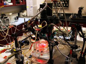
Researchers at The Ohio State University Wexner Medical Center developed a technique to revive parts of donated human hearts in the laboratory to search for hidden sources of irregular heartbeats. Submitted photo.
COLUMBUS, Ohio – Researchers have developed a technique that allows them to revive parts of human hearts in the laboratory for up to 12 hours while they search for hidden sources of irregular heartbeats.
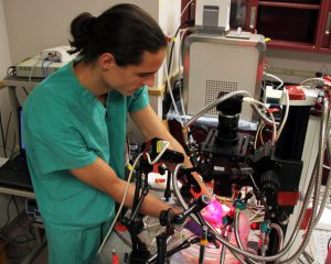
“It’s amazing. You can see the heart start beating again, which gives us an opportunity to study it with high-speed, high-tech equipment,” said Vadim Fedorov, PhD, a biologist who developed the technique at The Ohio State University Wexner Medical Center. “This allows us to see the heart in 3D and search for sources of irregular beats that we’ve never been able to find.”
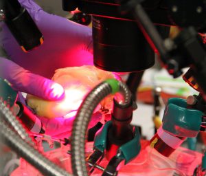
The hearts are donated by transplant patients who are receiving new hearts. Scientists take the top portions, known as the atria, and place them in a dish surrounded by four high-speed optical cameras. They then reanimate the tissue and inject it with a special dye that detects electrical signals.
“Normal imaging techniques give us one to two hundred recordings of the heart,” said Fedorov. “These cameras are able to give us 40,000 recordings, all in 3D.”
The images allow experts to more clearly map the heart and find new areas in which to perform ablations, a procedure during which tiny cuts or burns are made to form scar tissue on the heart. The scar tissue can help disrupt the electrical circuitry of the heart and stop irregular beats.
Knowing exactly where to ablate the small areas of the heart can be challenging, especially in patients who have persistent atrial fibrillation. Their hearts beat erratically 24 hours per day, which can lead to stroke or heart failure.
“This will allow us to map the heart more completely and eventually may help us personalize our procedures according to each patient’s individual needs,” said Dr. John Hummel, director of clinical electrophysiology research at Ohio State Wexner Medical Center. “While it’s still in the early stages, the work Dr. Fedorov is doing with human heart tissue is the kind of thing that will make pivotal changes in our ability to manage patients’ diseases in the coming years.”